Medical Imaging/ Radiology
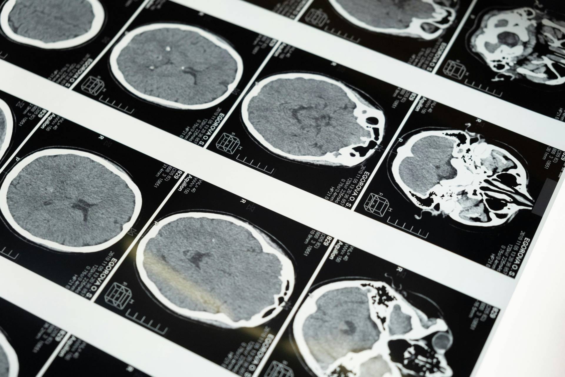
A CT scan combines a series of X-ray images taken from different angles to create cross-sectional “slices” of the bones, blood vessels, and soft tissues inside your body. It provides much more detail than a standard X-ray, making it an essential tool for diagnosing internal injuries, tumors, or complex bone fractures. The procedure is painless, non-invasive, and very fast, usually taking only a few minutes. It is often used in emergencies to quickly identify internal issues. Our advanced scanning technology ensures high-quality imaging with a focus on patient safety and accuracy.
An X-ray is a quick, painless imaging test that has been a cornerstone of medical diagnosis for decades. It is primarily used to visualize the body’s internal structures, particularly the skeletal system, to detect fractures, infections, or signs of arthritis. Beyond bones, X-rays are also vital for checking the chest and lungs for signs of pneumonia or heart enlargement. The procedure takes only seconds and requires no recovery time. It is a fundamental first step in diagnosing many common medical concerns with speed and precision.
An ultrasound scan uses high-frequency sound waves—rather than radiation—to create real-time images of the inside of your body. It is widely known for monitoring fetal development during pregnancy, but it is also an excellent tool for examining organs like the liver, gallbladder, and kidneys. Because it captures images in real-time, it can show the movement of internal organs and blood flowing through vessels. The process involves a handheld probe and a cooling gel, making it a completely safe and comfortable experience. It is a versatile diagnostic tool for patients of all ages.
An ECG is a simple, non-invasive test that records the electrical activity of your heart over a period of time. Small sensors called electrodes are placed on your chest and limbs to detect the electrical impulses that trigger every heartbeat. This test is crucial for diagnosing irregular heart rhythms (arrhythmias), identifying the cause of chest pain, or monitoring heart health after treatment. The procedure is entirely painless and usually takes less than ten minutes to complete. It provides an immediate “snapshot” of your heart’s performance.
Commonly known as a “cardiac ultrasound,” an echocardiography uses sound waves to create a detailed moving picture of your heart. This allows doctors to see the heart’s chambers, valves, and walls in motion, ensuring that the heart is pumping blood effectively. It is the primary tool for diagnosing valve problems, heart failure, or structural abnormalities without any surgery or radiation. The test is performed by moving a transducer over your chest, providing real-time data on your cardiovascular health. It is a vital, stress-free procedure for maintaining a healthy heart.
A Transcranial Doppler (TCD) is a specialized ultrasound that measures the velocity of blood flow through the brain’s blood vessels. It is a painless, non-invasive test used to identify issues like narrowings, blockages, or spasms in the arteries that supply the brain. TCD is particularly helpful in assessing the risk of stroke or monitoring conditions like sickle cell disease and brain trauma. By using sound waves to “hear” the blood flow, specialists can gain critical insights into your neurological health. The procedure is performed at the bedside and involves no radiation or injections.
A Renal Artery Doppler is a targeted ultrasound used to evaluate the blood flow to your kidneys. It specifically looks for any narrowing or blockages in the renal arteries, which can lead to high blood pressure or kidney dysfunction. This non-invasive test is an excellent way to check for a condition called renal artery stenosis without the need for needles or contrast dyes. By monitoring how well blood is reaching these vital organs, your doctor can better manage hypertension and overall kidney health. It is a comfortable, safe procedure that provides essential information for long-term wellness.
An endoscopy is a minimally invasive procedure that allows doctors to view the internal lining of your digestive tract using a thin, flexible tube with a camera. An Upper Endoscopy focuses on the esophagus, stomach, and small intestine, while a Lower Endoscopy (Colonoscopy) examines the colon and rectum. These procedures are essential for diagnosing digestive issues, identifying inflammation, or screening for polyps and cancer. Most patients receive light sedation to ensure the process is comfortable and quick. It is a vital tool for preventative health and precise diagnosis.
An HSG is a specialized X-ray procedure primarily used to evaluate female fertility and reproductive health. During the test, a gentle dye is introduced into the uterus and fallopian tubes to help them show up clearly on imaging. This allows specialists to check for any blockages in the tubes or structural issues within the uterus that might affect conception. The procedure is relatively brief and is usually performed in the first half of the menstrual cycle. It provides critical insights for patients on their journey toward reproductive wellness.
A urethrogram is a diagnostic imaging study used to examine the health and structure of the urethra. By using a safe contrast dye and X-ray technology, medical professionals can identify any narrowing (strictures), blockages, or injuries that may be causing urinary difficulties. This procedure is instrumental in diagnosing conditions that affect the flow of urine and helps in planning effective treatments or surgeries. It is a focused, efficient test conducted with the utmost care for patient comfort. Our team ensures a private and professional environment throughout the process.
A mammogram is a low-dose X-ray of the breast and is considered the “gold standard” for early breast cancer detection. This screening can identify changes or small lumps that are too tiny to be felt during a physical exam, often years before symptoms appear. Regular mammograms are a proactive step in women’s health, significantly increasing the success rate of early intervention and treatment. The process is quick, and our technicians are trained to make the experience as comfortable and supportive as possible. Taking twenty minutes for this scan provides invaluable peace of mind.